
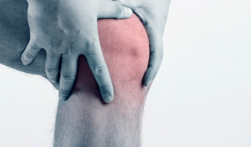
Regaining knee stability with physiotherapy
 The anterior cruciate ligament (ACL) is a fundamental part of the knee's anatomy, playing a crucial role in stabilizing the joint. However, ACL injuries can occur as a result of sports injuries, falls, or other accidents. The good news is that physical therapy can be a key element in the recovery process, helping patients regain knee function and return to daily activities and sports.
Importance of the anterior cruciate ligament (ACL)
 The ACL is one of the four main ligaments in the knee and its function is to prevent excessive anterior slippage of the tibia on the femur and to limit internal rotation of the tibia. It is responsible for preventing excessive movement between the tibia and femur. This ligament is particularly important for knee joint stability during activities that require changes of direction, such as in sports like soccer and skiing, where the knee is subjected to heavy stress. An ACL injury, in the acute phase, can cause instability, pain, and functional limitation of the joint, thus impairing the ability to participate in daily activities. However, the cruciate ligaments are not essential for proper gait; the truly important parameter is joint stability, which is essential during athletic activity.
Common causes of ACL injuries
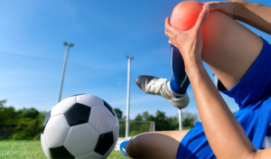 ACL injuries most often occur in sports situations involving strong twisting movements or sudden changes in direction. Sports injuries, such as soccer, basketball, and skiing, are often associated with ACL injuries. However, they can also occur in non-sports situations, such as falls or car accidents.
Rehabilitation phases following anterior cruciate ligament reconstruction
ACL rehabilitation is a complex process that requires time and commitment from the patient. Common rehabilitation phases include:
Reduction of pain, inflammation and swelling: after ACL surgery, swelling is often present, 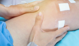 Redness, pain, and decreased range of motion. The physical therapist's initial goal is to reduce these common symptoms in the acute phase, restoring the patient to as normal a condition as possible. This process can sometimes take a month, partly because the initial phase is the most delicate for the patient, both emotionally and in terms of pain perception. The physical therapist can use swelling reduction techniques to improve the patient's comfort.
ROM Recovery(range of motion) joint, the range of motion: once the most difficult phase has been overcome, it will be necessary 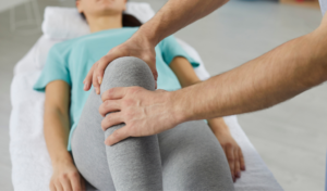 Recover full range of motion in the knee joint. Since the joint requires maximum range of motion to function properly, it is crucial not to leave it incomplete if you want to avoid future discomfort or problems. This second process requires an additional month. The patient will work with a physical therapist to restore full range of motion to the knee. This may include stretching and joint mobilization exercises.
Muscle strengthening: although muscle strengthening must begin from the first day after surgery, it will arrive on 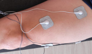 This is when you need to focus on strength. Following any surgical procedure, there is an immediate loss of weight and underlying muscle tone. Electrical stimulation and muscle recruitment must be performed from day one. Good exercise is like medicine: it soothes pain and reduces inflammation, as it stimulates catabolites, oxygenates the tissue through improved blood circulation, and consequently creates an immediate feeling of well-being in the patient.
The expression "it doesn't take much to feel good" can be applied to simple muscle contraction exercises that reduce pain. Therefore, once you reach the third phase of rehabilitation, you'll need to focus on strength and stability, maximally activating those weakened muscles, which are essential for a return to a sporty and active lifestyle. This process will take another month. Strengthening the muscles surrounding the knee is essential for restoring stability. Strengthening exercises will be an integral part of the rehabilitation program.
Return to functionality: the physiotherapist will guide the patient through specific exercises to improve stability and 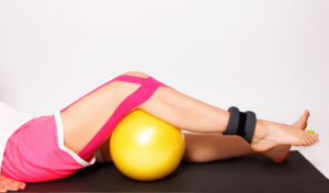 coordination, preparing him for the return to sports or daily activities (another month).
ACL rehabilitation requires patience and dedication, but the results can be very rewarding. Physical therapy is an essential part of this process, helping patients regain knee stability and live an active, healthy life.
Injury Prevention
Given the very high rate of recurrences on both the operated limb and the contralateral leg, it is essential to guide the patient towards continuous physical care for the following months. The total program should last between 6 and 9 months, starting with a physiotherapist and ending with specialized personnel, who must also understand the patient's needs and habits. It is obvious that a physiotherapist will require much more consistent support because they will be exposed to multiple stimuli, unlike those with a less dynamic routine. Preventing future injuries is an important aspect of ACL rehabilitation. The physiotherapist will provide advice on how to avoid similar injuries.
 In conclusion, recovery from an ACL injury requires a solid rehabilitation plan guided by an experienced physical therapist. Through targeted exercises and careful care, patients can return to enjoying the activities they love, minimizing the risk of future injuries. If you have suffered an ACL injury, consult a physiotherapist to begin the rehabilitation and recovery process.
Remember that every patient is unique, so it's important to work with a physical therapy professional to develop a personalized rehabilitation plan that fits your specific needs.


Via Donatello 75
00195 Rome
Tel. (+39) 06 39375719


Mon. – Fri. 8:00 – 20:00
Saturday 8:00 – 14:00

Via Donatello 75
00195 Rome
Tel. (+39) 06 39375719
Mon. – Fri. 8:00 – 20:00
Saturday 8:00 – 14:00

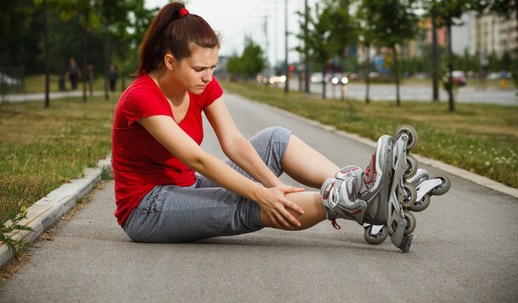
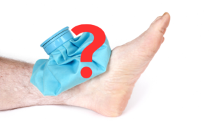 pain and swelling? The use of ice creates many doubtsFor example, it has been shown that internal temperature does not change, so endogenous cold does not pass through. Prolonged use can lead to a loss of responsiveness at the receptor level, resulting in a structure less responsive to external stimuli.
 leaking after the injury. Swelling is one of the post-traumatic effects, and to promote venous return, we must implement easily reproducible practices.
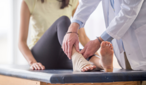 Make sure they are applied properly and sensibly. A poorly applied bandage can be worse than no bandage at all, so carefully examine the injury and assess the patient's needs.
 All these are important guidelines that we can recommend to the patient who has injured himself, but another highly recommended approach is to to be seen by a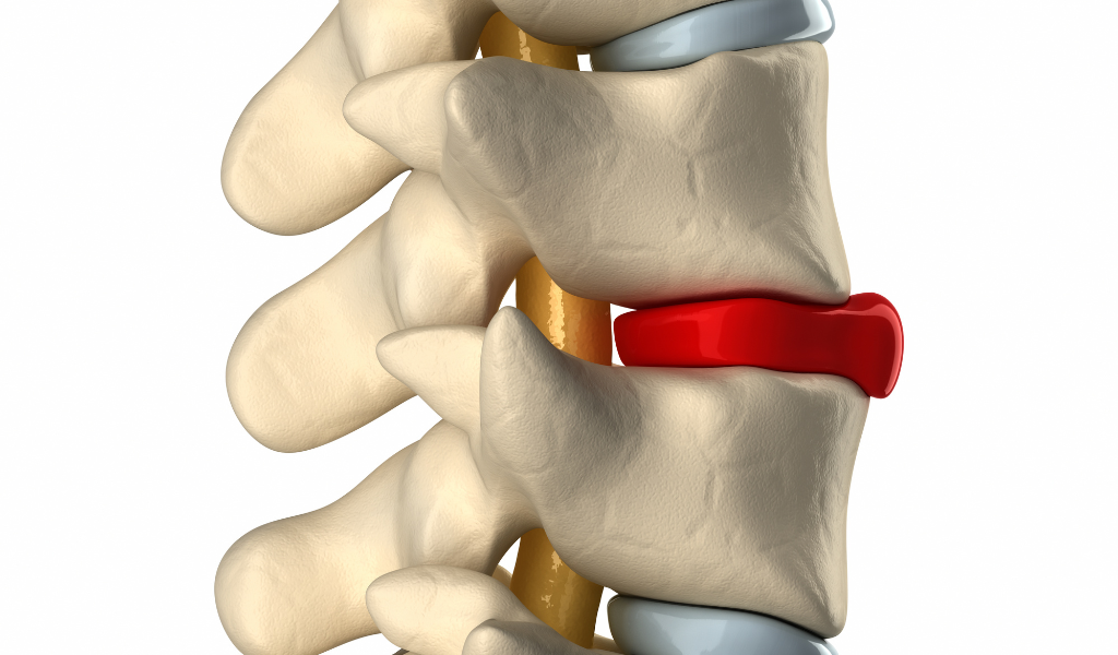
 The intervertebral disc is stretched and therefore taut, while the posterior wall is shortened and therefore relaxed. This condition could lead to the disc's only escape route being a posterior direction, contrary to what was claimed in the previous model.
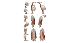 The nerves assume a more vertical direction, and the intervertebral foramen (TP3T) increases its area, creating more space for the structures within it. During extension, however, the spinal cord shortens and narrows the TP3T foramen. Although the nerve roots are relaxed during extension, the narrowing of the foramen, combined with a possible backward displacement of the intervertebral disc, could cause nerve compression. Clinical presentations are varied, and mixed components may occur, so the patient experiences pain during both flexion and extension.
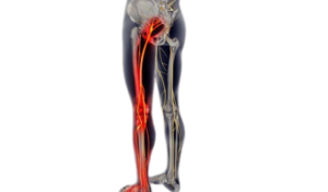 A very common example is the lumbosciatica, following the closure of the L5-S1 foramen (the smallest of the intervertebral foramina). If the herniation compresses the root, creating a radiculopathy, nerve conduction is blocked in the spinal nerve axon or its roots, causing hypoesthesia (numbness), muscle weakness, and/or reduced reflexes, but it is important to specify that this is not radicular pain. The association between hernia and pain is not correct, because we could only have silent symptoms., or have associated neuropathic pain, distributed across the dermatomes of the body, radiating below the knee and often to the foot. Therefore, the distribution of symptoms is very important; if they were to be anterior, it would be called lumbocruralgia, along the course of the crural nerve (L4 and proximal roots) instead the posterior distribution, the famous lumbosciatica, along the course of the sciatic nerve (L5-S1).
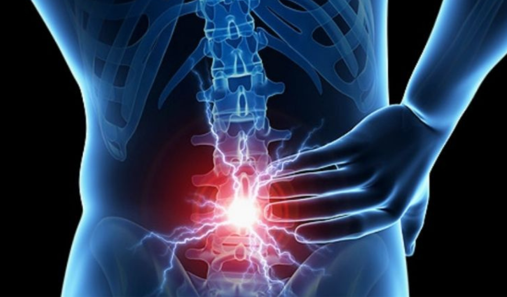 Colopathy is characterized by impaired sensation, strength, and reflexes, as well as the absence of pain. This condition is even more evident in chronic cases, where the symptoms are very severe but painless. This detail must always be taken into consideration, because the absence of pain does not indicate patient improvement. The patient's progress can only be considered evident in terms of regained sensation, strength, and reflexes. This assessment should be performed bilaterally to compare the affected side with the contralateral side.